|
Choroidal Nevus
Common “choroidal nevus” occurs in about 5 to 10
percent of the population. A nevus, freckle, or
mole can occur in the eye just like one can
occur on the skin. Like the skin, the eye has
melanocytes. Those are the cells that give the
skin its pigment. The eye has a layer of
melanocytes behind the retina in the choroid. If
a number of melanocytes grow, they can form a
nevus in the eye. These can only be seen by an
eye care expert who has the equipment and
the skill necessary to view the back of the eye.
A dilated examination is almost always
necessary. Like a nevus on the skin, a choroidal
nevus can grow into a malignant melanoma. |
|
 |
|
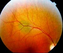
A choroidal nevus rarely
requires treatment. Photography is typically
used to document the size of the choroidal
nevus. If the choroidal nevus has orange
pigmentation, if the nevus is leaking fluid, or
has a thickness of 2 mm or more, it may be (or
become) a malignant choroidal melanoma.
Depending on its appearance, patients with a
choroidal nevus should have their eyes examined
every year. Currently, only your eye doctor can
look inside your eye to see if the choroidal
nevus has changed. If the choroidal nevus has
orange pigment or has thickened, it should be
checked more often. If a choroidal nevus is
leaking subretinal fluid, this is a particularly
ominous sign. Such nevi should be followed most
closely for evidence of growth or malignant
transformation into a choroidal melanoma. The
risk of a choroidal nevus transforming into a
choroidal melanoma is about 1 in 20,000.
Choroidal nevus is typically a “pigmented tumor”
of the blood vessel layer (choroid) beneath the
retina. A choroidal nevus is typically gray but
can be brown, yellow, or variably pigmented.
Your eye care professional will look to see if
the choroidal nevus is raised (has thickness),
has orange pigment (lipofuscin), or is leaking
fluid (retinal detachment). If the choroidal
nevus has one or more of these finding, it is
labeled a suspicious choroidal nevus that might
turn into (or be) a small choroidal melanoma.
If the choroidal nevus looks suspicious, it is
reasonable to have an eye cancer expert
check it. This examination may include the use
of ultrasound, concentrated photography, or an
intraocular angiogram. It is a good idea to keep
a picture of your choroidal nevus. This picture
can be compared to future examinations to help
determine if the nevus has changed or stayed the
same.
A choroidal nevus can have yellow-white spots on
its surface called drusen. This is a sign that
the choroidal nevus is preventing the eye from
removing retinal waste products. It is also a
sign that the choroidal nevus has been present
for enough time for these products to
accumulate. There are no studies that show how
long it takes for drusen to form on a choroidal
nevus.
A benign choroidal nevus requires no treatment
and there is no way to safely remove them. Since
a choroidal nevus can turn into a choroidal
melanoma, it is reasonable to have it
periodically observed by your eye care
professional. Since skin and conjunctival
melanomas have been linked to ultraviolet
exposure, and since choroidal melanomas are more
commonly found in patients with blue eyes, those
with outdoor occupations, and those living in
Australia (where there is an ozone hole), it is
reasonable to wear ultraviolet (UV) blocking
sunglasses. Think of sunglasses as “sun block
for your eyes”. |